Researchers at Weill Cornell Medicine and Cornell Engineering have utilized state-of-the-art tissue engineering techniques and a 3D printer to construct a replica of an adult human ear that closely mimics the look and feel of a natural ear. The study, which was published online in Acta Biomaterialia on March 16, holds the potential to provide grafts with precise anatomical features and appropriate biomechanical properties for individuals born with congenital ear malformations or those who have lost an ear later in life.
Dr. Jason Spector, chief of the Division of Plastic and Reconstructive Surgery at NewYork-Presbyterian/Weill Cornell Medical Center and a professor of surgery (plastic surgery) at Weill Cornell Medicine, highlighted the challenges of ear reconstruction, emphasizing the need for multiple surgeries and a high level of skill and precision. He expressed hope that this innovative technology could offer a realistic option for the thousands of individuals requiring surgery to correct outer ear deformities.
Traditionally, surgeons have relied on harvesting cartilage from a child's ribs to construct a replacement ear, a procedure that can cause pain and scarring. Despite efforts to shape the graft to resemble the recipient's natural ear, it often lacks the same flexibility and realism.
One way to produce a more natural replacement ear is to enlist the aid of chondrocytes, the cells that build cartilage. In earlier studies, Dr. Spector and his colleagues used animal-derived chondrocytes to seed a scaffold made of collagen, a key component of cartilage. Though these grafts developed successfully at first, over time the well-defined topography of the ear—its familiar ridges, curves, and whorls—were lost. "Because the cells tug on the woven matrix of proteins as they labor, the ear contracted and shrank by half," said Dr. Spector.
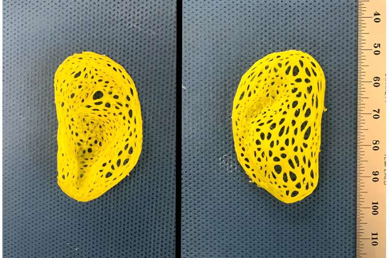
To address this problem in this study, Dr. Spector and his team used sterilized animal-derived cartilage treated to remove anything that could trigger immune rejection. This was loaded into intricate, ear-shaped plastic scaffolds that were created on a 3D printer based on data from a person's ear. The small pieces of cartilage act as internal reinforcements to induce new tissue formation within the scaffold. Much like rebar, it strengthens the graft and prevents contraction.
Over the next three to six months, the structure developed into cartilage containing tissue that closely replicated the ear's anatomical features, including the helical rim, the "anti-helix" rim-inside-the-rim, and the central, conchal bowl. "That's something that we had not achieved before," said Dr. Spector.
To test the feel of the ear, biomechanical studies were performed in conjunction with Dr. Spector's longtime engineering collaborator Dr. Larry Bonassar, the Daljit S. and Elaine Sarkaria Professor in Biomedical Engineering at the Meinig School of Biomedical Engineering on Cornell's Ithaca campus. This confirmed that the replicas had flexibility and elasticity similar to human ear cartilage. However, the engineered material was not as strong as natural cartilage and could tear.
To remedy this issue, Dr. Spector plans to add chondrocytes to the mix, ideally ones derived from a small piece of cartilage removed from the recipient's other ear. Those cells would lay down the elastic proteins that make ear cartilage so robust, producing a graft that would be biomechanically much more similar to the native ear, he said.


0 Comments