In 1977, recombinant protein expression revolutionized biology, but screening protein clones remained an intricate and exhausting task. During a sabbatical at Duke University, George Smith, a biochemist from the University of Missouri, began pondering how he might simplify this cumbersome process. His research had long focused on bacteriophages, and he wondered if a specific coat protein, pIII, from the filamentous bacteriophage he studied could be adapted for this purpose. With this idea in mind, he teamed up with Duke University biochemists Robert Webster and Paul Modrich, and together, they devised an innovative approach that would become known as "phage display."
The team’s breakthrough introduced a method they could barely anticipate the full potential of—a form of "simple evolution in a Petri dish," as Smith would later describe it in his Nobel lecture. Yet, questions persisted. What hidden complexities lay within this newly discovered mechanism? As they celebrated their initial success, doubts nagged: Could phage display truly be harnessed to mimic natural selection, or were there unknown limitations that might arise? How many more applications could this technique unlock? Their collaborative spirit mixed with a touch of trepidation, as each experiment unearthed deeper mysteries and glimpsed at the possibility of entirely new fields yet to be uncovered.
In the early and mid-1980s, scientists were exploring ways to use bacteriophages to express recombinant proteins, a revolutionary idea at the time. The method they initially developed involved painstakingly isolating viral plaques on Petri plates and transferring them onto membranes, then treating these membranes with antibodies to locate desired proteins. This process, though groundbreaking, was complex and time-consuming, requiring careful mapping of each antibody signal back to the original viral plaque—a task fraught with opportunities for error and inefficiency.
Then came Smith, who had a bold new vision to streamline and accelerate this screening process. He proposed attaching foreign proteins to one end of the phage coat protein, pIII, allowing scientists to select for these modified phages directly using antibodies bound to surfaces. This approach promised a transformative leap: by eliminating unnecessary clones, scientists could focus solely on phages with the desired protein, massively increasing screening efficiency. Yet, even with this breakthrough, questions loomed. Could this method handle more complex proteins without failure? Would the antibody selection prove too restrictive, potentially discarding valuable variants? As researchers adopted Smith’s approach, they were both excited and apprehensive, confronting these mysteries with a blend of optimism and caution. Each day, they wondered if they were on the cusp of something monumental—or if unforeseen limitations lay ahead.
When the team first realized that they could link a specific genotype to its corresponding phenotype by binding a protein to bacteriophages carrying inserted DNA, it opened up an array of thrilling possibilities—and mysteries. The groundbreaking concept allowed scientists not only to acquire a specific phage clone but also to sequence it, unveiling both the nucleotide and amino acid sequence of peptides displayed on its surface. Jamie Scott, who had joined George Smith's lab in the late 1980s and later became a professor emeritus at Simon Fraser University, described the process as revolutionary. The potential seemed boundless, but as Smith returned to his lab, he found that deeper questions lay ahead.
In 1985, Smith published his pioneering research on "filamentous phage display," revealing a method that allowed for screening billions of peptides simultaneously. However, this approach left some unanswered questions. What if some amino acids were overrepresented due to redundancy in genetic coding? Could they control amino acid frequencies, or would certain residues dominate randomly generated sequences? The randomness posed a dilemma—how could they ensure that the best peptides were identified without control over amino acid variations? As later researchers worked to synthesize specific codons with precise characteristics, the team faced an even deeper enigma: would these carefully curated libraries truly enhance binding affinities, or would they encounter new unforeseen limitations? The answers, they knew, could redefine the future of molecular biology.
“It’s not random,” said Anthony Kossiakoff, a protein engineer at the University of Chicago who delves into the intricate relationships between protein structures and their functions. “There’s a lot of planning and designing for how you can best utilize what you’ve got.” His words lingered in the minds of his colleagues, as he highlighted the hidden precision behind a process that might otherwise seem arbitrary. For Kossiakoff and other structural biologists, the journey to high-affinity protein selection is filled with complex layers. They must not only consider their vast libraries of proteins but also the fundamental nature of the antigens themselves. “The biggest factor,” Kossiakoff noted, “is the quality of the antigen.” But what exactly defines this quality?
Questions proliferate as researchers wrestle with unknowns: Is the antigen stable enough to yield a usable result? How can they confirm it maintains the desired conformation throughout the selection process? To secure high-affinity binding, teams must impose strict conditions in the affinity purification steps, adjusting factors such as ligand concentration or even competing enzymes. Yet, as Kossiakoff hinted, even with careful planning, each decision is a gamble. And when the final genotype-phenotype linkage is achieved, what secrets remain hidden within the extracted genetic sequences?
**Opening the Door**
The connection between tailored instructions and the resulting bound product introduced an entirely new paradigm in the research world, transforming how scientists could screen vast numbers of candidates. This model was especially appealing for antibody research, which requires precise matching of antibodies to specific targets, and soon became a staple in labs worldwide. But despite the potential, questions lingered. Why did some peptide sequences bind more effectively than others? And was there an underlying pattern yet to be discovered within these vast combinations?
While George Smith’s original method created random peptide sequences and tested their fit against known antibodies, other scientists, like John McCafferty, wondered if the process could be reversed. “What if we could design antibodies directly against specific targets?” he asked, captivated by the possibilities this approach suggested. Together with his mentor, Greg Winter, McCafferty began exploring phage display technology to test this hypothesis.
As they worked, new questions emerged: Could this technique allow for more personalized treatments? What unknowns lurked within the thousands of tested sequences? The scientists couldn’t help but feel a mounting tension, as each breakthrough revealed yet another layer of mystery. Unanswered questions hinted at hidden complexities, suggesting that the research had only scratched the surface of what might be possible.
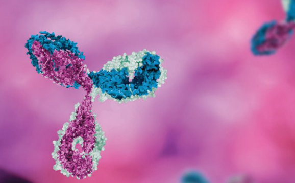
The groundbreaking development of single-chain Fv (scFv) technology transformed antibody engineering, yet it raised as many questions as it answered. By cloning the variable regions of the heavy and light chains of antibodies from immunized blood donors into a single coding region, researchers McCafferty and Winter succeeded in creating a fusion protein that retained full antigen-binding properties—essentially a compact version of a complete antibody. They called this creation the single-chain Fv (scFv), which could then be expressed by bacteriophages. Remarkably, these scFvs could be affinity-selected in a manner similar to Smith's peptide selections, using an immobilized target peptide to isolate antibodies with precise binding capabilities.
Despite the success, a host of mysteries remained. McCafferty admitted, "A feat that I've tried to repeat ever since without success," hinting at the difficulties in recreating this innovation. Why were some scFvs easier to produce than others? And what factors influenced their binding specificity? Compared to the traditional hybridoma technique, which involved labor-intensive processes and months of waiting, phage display promised speed and efficiency by allowing scientists to amplify genetic material from B cells in bacteriophages almost instantly. But what unforeseen challenges might accompany this new method? Could phage display unlock antibodies that could not only bind effectively but also avoid immune rejection in humans? And while hybridomas required time-consuming "humanization" of animal antibodies, phage display’s potential for fully human antibodies opened up new therapeutic possibilities. This leap forward left scientists and clinicians both excited and cautious, wondering what boundaries—if any—might remain in their search for novel treatments.
This groundbreaking method gave rise to the world’s first antibody created through phage display technology—the anti-TNF antibody adalimumab, widely recognized as Humira. This scientific breakthrough was achieved by Cambridge Antibody Technology, the company co-founded by McCafferty and Winter, with crucial support from the chemical giant BASF. Yet, the journey to Humira’s development was riddled with unanswered questions and unexplored territory, leaving even the scientists wondering just how far phage display could go in antibody engineering.
As researchers pushed the boundaries, a new frontier opened with next-generation sequencing paired with phage display, allowing for an unparalleled speed in producing and analyzing antibodies. But what are the true limits of this technology? Some targets previously considered unreachable now seemed within grasp, yet mysteries linger. What hidden risks might arise as they create antibodies with no “basis for tolerance” in synthetic libraries, as Scott noted? Could there be unforeseen consequences when synthetic antibodies enter a complex biological system? For the scientists, the possibilities were as thrilling as they were daunting, each discovery raising new questions. How much control did they truly have over this powerful process, and what would happen if the technology advanced beyond their ability to predict its impact?
Phage display is a formidable and evolving technique in the field of antibody production. This technology, initially relying on bacteriophages to present antibody fragments, has now expanded to incorporate a range of alternative display vehicles, including unconventional viruses, various bacterial strains, and even eukaryotic cells like yeast and mammalian cell lines. Yet, as Dr. McCafferty notes, phage display remains somewhat enigmatic. Researchers often work with large, complex libraries and intricate antigen interactions, but what truly happens at each stage remains partly mysterious. After running a display experiment, scientists may be left with a vast number of potential binding candidates—many more than they can feasibly assess in detail for binding affinity or cross-reactivity with other targets. This leaves questions: How many of these interactions are meaningful, and could some go undetected due to experimental limitations?
In addition to phages, yeast and mammalian cell displays, though limited in library size, offer compatibility with flow cytometry and cell-sorting techniques, enhancing researchers' ability to examine protein clones’ specific biophysical traits. There’s also a growing interest in exploring alternative molecular backbones beyond immunoglobulin, such as protein domains like the fibronectin-binding domain, to overcome structural limitations of traditional antibodies. As the boundaries of phage display broaden, researchers are left wondering about the vast, uncharted potential of these innovative approaches, as well as the hidden complexities that may lie beneath each experimental success.
Recent advancements in modified selection methods have greatly expanded the ability to identify proteins with specific, desirable characteristics. For instance, in *in vivo* selection, candidate proteins are delivered directly into model organisms, where scientists can observe their distribution across various organs. This technique raises a series of intriguing questions: why do certain proteins target specific organs while others do not? What mechanisms govern this selective distribution, and could it reveal hidden pathways within the body?
In addition to *in vivo* testing, researchers use whole-cell culture methods to screen protein libraries across multiple cell types, exploring how different receptors react to each protein. Here too, the unknowns multiply—what drives certain proteins to bind to specific receptors, and could these interactions suggest undiscovered cellular functions or vulnerabilities?
Beyond antibodies, phage display techniques are employed to examine protein interactions at an atomic level, revealing the evolutionary forces that have shaped these molecular connections. As Dr. Kossiakoff explains, these methods let researchers probe complex questions about protein interface energetics, uncovering which aspects are evolutionarily preserved and why.
Phage display also shows promise in identifying rare or synthetic peptides with potential as cancer therapeutics, allergen identifiers, and even antivenom antibodies. Yet, many mysteries remain: could these peptides be harnessed to treat diseases beyond cancer, or even neutralize toxins from multiple venomous species? The possibilities seem boundless, yet the answers remain tantalizingly out of reach, leaving scientists on the brink of discovery.


0 Comments